
Intra-Articular Combination of Low-Molecular-Weight Collagen and a Corticosteroid in Advanced Knee Osteoarthritis: A Case Report
Authors: Milan Rejholec1,2*
- 1Centrum Prof. Čecha s.r.o., Prague, Czech Republic
- 22nd Orthopedic Clinic, University Hospital Motol and Homolka, Prague, Czech Republic
- * corresponding author
- Journal
- Clinical Pathology & Research Journal · Volume 10(1): 000224
- Published
- Received
- ISSN
- 2642-6145
Version
Original English text of the study.
Source document
On this page
Key findings
- A 76-year-old patient with bilateral KL grade III knee osteoarthritis received PEPTYS with betamethasone after previous conservative treatment had failed.
- At one year, pain fell from 8/10 to 2/10, effusion resolved, flexion improved by 5° in both knees and regular analgesic use stopped.
- A single case and the corticosteroid co-intervention cannot isolate the effect of collagen or prove cartilage regeneration.
- patient
- 1
- combined treatment administrations
- 5
- VAS pain (0-10)
- 8 → 2
- months of follow-up
- 12
Advanced knee osteoarthritis (Kellgren–Lawrence [KL] grade III–IV) markedly impairs quality of life and is a common indication for total knee arthroplasty (TKA). In elderly, polymorbid patients, or when surgery is contraindicated, minimally invasive conservative options become important. We report a 76-year-old man with advanced bilateral knee osteoarthritis (KL grade III, more severe on the left) in whom TKA was contraindicated because of a history of lower-limb erysipelas. After non-steroidal anti-inflammatory drugs (NSAIDs), symptomatic slow-acting drugs for osteoarthritis (SYSADOAs), repeated joint aspiration and intra-articular hyaluronic acid had failed to provide lasting relief, intra-articular therapy combining low-molecular-weight collagen peptides (PEPTYS, 2 mL) with a corticosteroid (betamethasone) was started. Five injections were given: the first three at three-month intervals and the last two at monthly intervals. One year after starting therapy, pain on a visual analogue scale (VAS) had fallen from 8/10 to 2/10, the recurrent joint effusion had resolved, knee flexion had improved bilaterally, and the patient had stopped all regular analgesics. Follow-up standing weight-bearing radiographs showed an apparent widening of the medial joint space on the right. No local or systemic adverse effects occurred. In selected polymorbid patients with advanced knee osteoarthritis and a contraindication to surgery, this intra-articular combination relieved symptoms and postponed arthroplasty. As this is a single case with a corticosteroid co-intervention, the symptomatic contribution of the collagen component cannot be isolated and controlled studies are warranted.
Knee osteoarthritis (OA) is a progressive degenerative disease affecting all structural components of the joint. In advanced stages (KL grade III–IV 1) there is severe loss of hyaline cartilage, subchondral sclerosis and reactive synovitis, presenting clinically with swelling, stiffness and pain at rest and on exertion. The concentration of hyaluronic acid in synovial fluid falls in parallel with the severity of structural damage 2. Although joint replacement (TKA) is the standard treatment for these stages, operative risk and comorbidity must be weighed in elderly and polymorbid patients; international recommendations (Osteoarthritis Research Society International, OARSI) therefore emphasise the role of minimally invasive conservative treatment, including intra-articular injections, in this group 3.
Alongside viscosupplementation with hyaluronic acid and autologous platelet-rich plasma, intra-articular injection of collagen is increasingly used in conservative management 4. PEPTYS, registered as a medical device, is a solution of low-molecular-weight collagen peptides (molecular weight < 3 kDa) obtained by targeted hydrolysis of collagen [5 ]; it belongs to the same class of hydrolysed collagen (< 3 kDa) for which intra-articular use has been clinically evaluated 6,7. The proposed mechanism is direct support of the extracellular matrix, down-regulation of inflammatory cytokines and stimulation of endogenous synthesis of hyaluronic acid and collagen by chondrocytes 6,8. We describe the long-term effect of this combined therapy in an elderly polymorbid patient.
A 76-year-old man presented to the orthopaedic outpatient clinic with long-standing, progressive pain in both knees, worse on the left. The pain occurred both on initiating movement and at rest and markedly limited walking on level ground (under about 200 m) and stair climbing. Previous conservative treatment — oral NSAIDs, SYSADOAs, repeated aspiration of both knees and intra-articular hyaluronic acid — had produced no lasting benefit. His medical history included two myocardial infarctions, radiotherapy for prostate cancer, and erysipelas of the lower limbs at the age of 70. In his youth he had undergone arthroscopy of the left knee twice and had played competitive football.
On examination there was bilateral varus deformity, tenderness over the medial joint line on the left, joint swelling without a large effusion, and restricted range of motion (flexion 95° on the left and 105° on the right, with a 5° extension deficit bilaterally). Baseline pain was rated 8/10 on the VAS. Ultrasound of both knees showed irregularity of the articular surfaces at the medial and lateral margins of the distal femoral condyles and proximal tibia bilaterally.
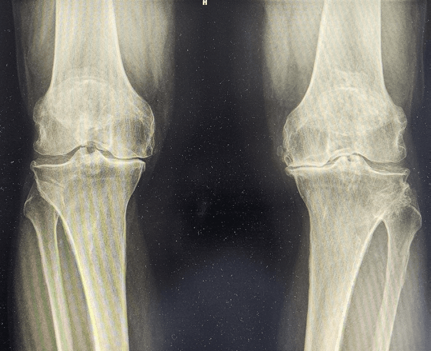
Standing weight-bearing radiographs showed advanced degenerative change: marked joint-space narrowing (predominantly medial), more severe on the left where the medial space was almost obliterated, with subchondral sclerosis, cysts and marginal osteophytes. The findings were graded as KL grade III bilaterally, with greater functional and structural impairment on the left 1.
After three injections of a leucocyte-poor platelet-rich plasma and hyaluronic acid preparation (Cellular Matrix) at 14-day intervals into both knees, symptoms on the left improved while the right knee remained symptomatic. A specialist centre recommended TKA of the right knee; the patient returned 6 months later reporting that surgery had been contraindicated because of his history of skin infection (erysipelas). At the patient’s request, hyaluronic acid was then injected into both knees four times at 4-month intervals, again without lasting benefit. Aspiration yielded clear, straw-coloured fluid — a total of 80 mL from the left and 47 mL from the right knee.
Given the patient’s age, comorbidity and wish to avoid surgery, intra-articular treatment was started combining the medical device PEPTYS (a 2 mL pre-filled syringe containing a solution of low-molecular-weight collagen peptides, < 3 kDa) 4 with the corticosteroid betamethasone (Diprophos, 7 mg/mL suspension) 8, to control the synovitis associated with the recurrent effusion. Five injections were given in total: the first three at three-month intervals (baseline, +3 and +6 months) and the next two at monthly intervals (+7 and +8 months); the interval was shortened in the second phase because of persistent intra-articular effusion. Over the series, a total of 90 mL was aspirated from the right and 167 mL from the left knee. Injections were performed in the outpatient setting using a standard anterolateral approach under strict aseptic conditions and were well tolerated, with no local or systemic adverse effects.
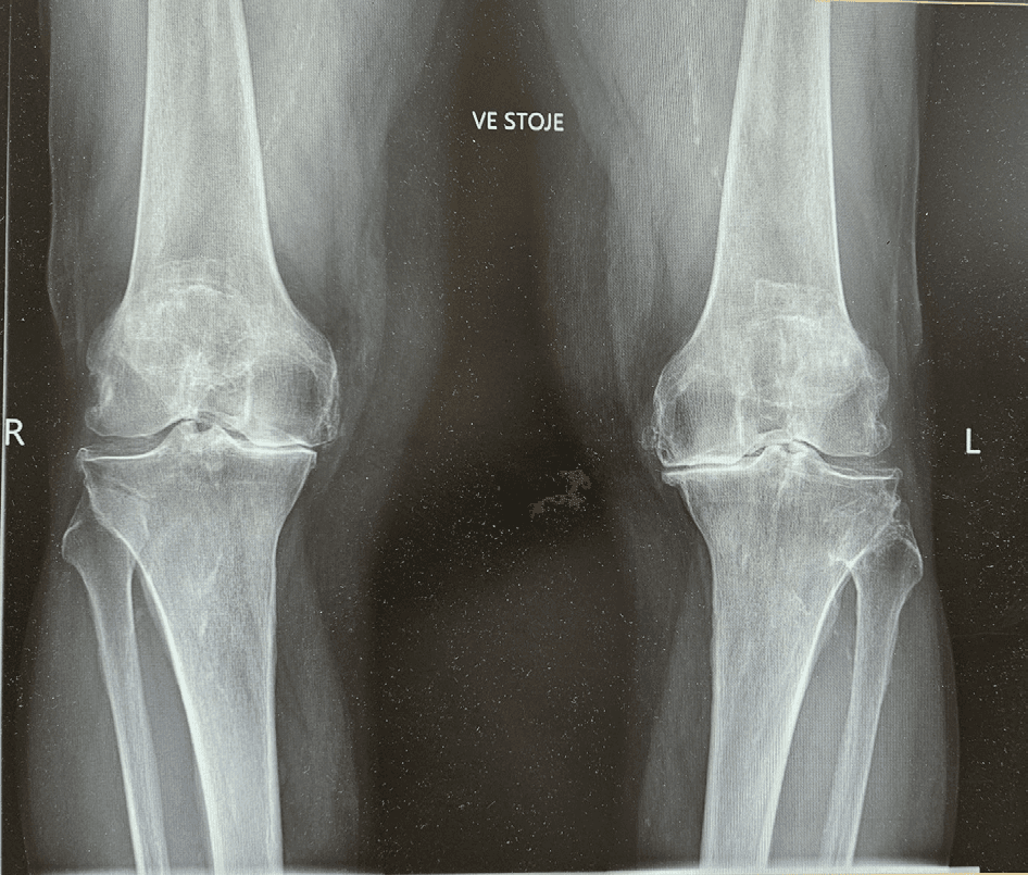
Clinical assessment was performed one year after the start of combined therapy. The patient reported a major reduction in pain at rest and at night; VAS pain fell from 8/10 to 2/10. Objectively, joint-line tenderness was reduced, knee flexion improved on both sides (right 105°→110°, left 95°→100°) and the pain-free walking distance increased. The effusion ceased and no further aspiration was required after the last injection, and the patient stopped all regular analgesics, including NSAIDs. This clinical improvement followed the start of the collagen injections. Comparison of the baseline weight-bearing radiograph (Figure 1; taken before the originally planned arthroplasty) with the follow- up weight-bearing radiograph (Figure 2) showed an apparent widening of the medial joint space on the right and less pronounced articular surface irregularity; both radiographs were obtained using the standard standing technique. The course of the clinical parameters is summarised in Table 1.
| Parameter | Baseline | One-year follow-up | Change |
|---|---|---|---|
| Pain (VAS 0-10) | 8/10 | 2/10 | -6 points |
| Flexion - right knee | 105° | 110° | +5° |
| Flexion - left knee | 95° | 100° | +5° |
| Joint effusion | Present (repeated aspiration) | None | Resolved |
| Analgesic use | Regular (daily) | None / occasional | NSAIDs stopped |
| Tolerance of treatment | - | Excellent | No local complications |
VAS = visual analogue scale; NSAIDs = non-steroidal anti-inflammatory drugs.
Treatment of advanced knee OA in elderly, polymorbid patients is a clinical challenge. With advanced cartilage loss (KL grade III), standard viscosupplementation with hyaluronic acid often fails because of the joint’s limited biological response. Low-molecular-weight collagen peptides offer a different mechanism: they penetrate the deeper extracellular matrix and, according to available work, act as signalling molecules that stimulate synoviocytes and chondrocytes to synthesise joint matrix while down-regulating the sterile inflammation induced by degraded tissue 3,7,9.
Intra-articular collagen has been associated with reduced pain, improved function and lower NSAID use. In a cohort of 309 patients, intra-articular polymerised type I collagen postponed the indication for TKA — most patients maintained clinical benefit over follow-up of up to 60 months and none required TKA 9. A pilot study by De Luca P , et al. of intra-articular hydrolysed collagen (< 3 kDa), the same class as PEPTYS, showed improvement in Western Ontario and McMaster Universities Osteoarthritis Index (WOMAC) scores without serious adverse effects 5, later supported by the larger multicentre study of Volpi p, et al. 7. A beneficial effect of type I collagen in painful knee OA was also reported by Stančík, et al. 10. A recent meta-analysis (35 randomised trials, 3165 patients) concluded that collagen derivatives are effective and safe in OA 11,12. The course in our patient is consistent with these data.
A feature of this case is the prolonged, intermittent dosing schedule. Unlike short-course regimens (e.g. 3–5 injections in quick succession), gradual administration stabilised the joint environment and resolved the chronic exudative synovitis, which in an older patient with slower tissue turnover allowed the indication for surgery to be averted.
The corticosteroid was used deliberately for rapid control of the marked synovitis and effusion; its anti-inflammatory and analgesic effect is rapid and, according to international recommendations, is appropriate for short-term pain relief in knee OA 3. The collagen peptides, by contrast, are credited mainly with stimulating synthesis of the joint’s own matrix 6,8. A purely symptomatic, anti-inflammatory mechanism would not be expected to alter the radiographic appearance; if anything, intra-articular corticosteroids are associated with concerns about cartilage rather than with any gain in joint space. In our patient the apparent widening of the medial joint space was seen on standard standing weight-bearing radiographs, and the clinical improvement followed the collagen injections. A single uncontrolled observation cannot prove cartilage regeneration, and a positioning artefact can never be entirely excluded; however, with a standardised weight-bearing technique a purely positional explanation is unlikely. This finding therefore argues, at least in part, against the view that the benefit was purely symptomatic and is compatible with a structural, matrix-stimulating contribution of the collagen component — a hypothesis that warrants confirmation in controlled studies.
This is a single case (n = 1) without a control group, so the findings cannot be generalised. Because the two agents were given together, the symptomatic benefit — particularly the early reduction in pain and effusion — cannot be separated from the effect of the corticosteroid. Assessment relied mainly on the VAS and range of motion; a validated functional score (e.g. WOMAC) was not used. The radiographs were standard standing weight-bearing views but were not quantitatively measured (for example with a fixed-flexion protocol), so the apparent change in joint space should be interpreted with caution and confirmed using standardised quantitative imaging. Controlled studies with objective, blinded outcomes are needed to establish the independent efficacy of low-molecular-weight collagen peptides.
In a polymorbid patient with advanced knee OA (KL grade III) and a contraindication to TKA, the intra-articular combination of low-molecular-weight collagen (PEPTYS) and a corticosteroid was associated with marked clinical improvement. The reduction in pain (VAS 8→2), resolution of effusion and improved range of motion increased the patient’s quality of life and allowed invasive surgery to be postponed. Given the nature of the report (a single case with combined treatment), controlled studies are needed to confirm the independent efficacy of the collagen peptides.
- Patient consent
- Written informed consent was obtained from the patient for publication of this case report and the accompanying radiographs.
- Conflict of interest
- The authors declare that they have no conflict of interest.
- Funding
- This work received no specific grant from any funding agency.
Transcribed from the supplied English PDF. The publisher’s promotional page is omitted; source reference numbering is retained. An editorial note flags the conflicting laterality in Figure 2. © 2026 Milan Rejholec. Source license: CC BY 4.0 (https://creativecommons.org/licenses/by/4.0/).
- 1.Kellgren JH, Lawrence JS (1957) Radiological Assessment of Osteo-Arthrosis. Ann Rheum Dis 16(4): 494-502.
- 2.Bannuru RR, Osani MC, Vaysbrot EE, Arden NK, Bennell K, et al. (2019) OARSI guidelines for the non-surgical management of knee, hip, and polyarticular osteoarthritis. Osteoarthritis Cartilage 27(11): 1578-1589.
- 3.Tarantino D, Mottola R, Palermi S, Sirico F, Corrado B, et al. (2023) Intra-articular collagen injections for osteoarthritis: a narrative review. Int J Environ Res Public Health 20(50): 4390.
- 4.PEPTYS-52 - Instructions for use (solution of low-molecular-weight collagen peptides <3 kDa) (2026). Manufacturer: TissYou Srl, Strada di Palerna, 2-47895 Domagnano (RSM); Distributor: Eumed S.r.l., Via N. Tommaseo, 68-35131 Padova, Italy.
- 5.De Luca P, Colombini A, Carimati G, Beggio M, Girolamo DL, et al. (2019) Intra-articular injection of hydrolyzed collagen to treat symptoms of knee osteoarthritis. A functional in vitro investigation and a pilot retrospective clinical study. J Clin Med 8(7): 975.
- 6.Volpi P, Zini R, Erschbaumer F, Beggio M, Busilacchi A, et al. (2021) Effectiveness of a novel hydrolyzed collagen formulation in treating patients with symptomatic knee osteoarthritis: a multicentric retrospective clinical study. Int Orthop 45: 375-380.
- 7.Carballeda FJ, Lima G, Llorente L, Alvarez NC, Ordaz RBH, et al. (2012) Polymerized-type I collagen downregulates inflammation and improves clinical outcomes in patients with symptomatic knee osteoarthritis following arthroscopic lavage: a randomized, double-blind, and placebo-controlled clinical trial. Scientific World Journal 2012: 342854.
- 8.Diprophos - Summary of product characteristics (betamethasone 7 mg/mL suspension for injection) (2017). Reg. no. SUKLS232371/2017. Prague: State Institute for Drug Control.
- 9.Borja-Flores A, Macías-Hernández SI, Hernández-Molina G, Perez-Ortiz A, Reyes-Martínez E, et al. (2020) Long-term effectiveness of polymerized-type I collagen intra-articular injections in patients with symptomatic knee osteoarthritis: clinical and radiographic evaluation in a cohort study. Adv Orthop 2020: 9398274.
- 10.Stančík R, Zvarka J, Hlaváč M, Kubinec V, Rovenský J (2012) Collagen type I in the treatment of painful osteoarthritis of the knee. Reumatologia 50(5): 390-395.
- 11.Liang CW, Cheng HY, Lee YH, Liao CD, Huang SW, et al. (2024) Efficacy and safety of collagen derivatives for osteoarthritis: a trial sequential meta-analysis. Osteoarthritis Cartilage 32(5): 574-584.
- 12.Kotaška J, Hanousková L, Průša R, Trč T, Kotaška K, et al. (2021) Concentration of hyaluronic acid in synovial fluid as an indicator of severity and degree of impairment in patients with gonarthrosis. Acta Chir Orthop Traumatol Cech 88(2): 83-86.
Citation
Rejholec M. Intra-Articular Combination of Low-Molecular-Weight Collagen and a Corticosteroid in Advanced Knee Osteoarthritis: A Case Report. Clin Pathol. 2026;10(1):000224. doi:10.23880/cprj-16000224.